Introduction:
For this lab I wanted to make a three dimensional structure to represent the complexity of a moving limb. It was not an easy task to build a model with all the major characteristics of an actual moving limb. So for my model I chose to take the concepts and simplify them as much as possible so they can be represented visually. My goal was to have a moving limb model that actually looked similar to a human limb, this makes it easier for the imagination to see the more complex parts of the limb.
Limb parts:
Humerus bone = plastic piping
Radius and Ulna bone = plastic piping
Bicep brachii and tricep brachii = paint roller cover (red)
Bundle of muscle fibers with thousands of myofibril = weather proofing insulation (white)
Elbow joint = small metal hinge
Neuron with dendrites = kids squishy toy (purple and teal)
Sensory Neuron (purple)
Motor Neuron (teal)
Axon = copper wiring
Myelin sheath = yellow wire clamps
Large muscle cell (sarcolemma) = blue pencil tip eraser
T-tubules = circular pencil grips (green)
Calcium = small round wooden balls
Sarcomere with single actin-myosin unit = adjustable metal device
Myosin with double-stranded DNA = Dark teal and purple pencil grips
 Here are the basic components I used to construct my movable limb. In this photo I have already cut the paint roller cover and started building the bicep and bundle of muscle fibers.
 I used a dremel to seperate the paint roller cover, I did not relize the inside was a strong plastic which turned out to be much stronger then I originally thought.
 This is a good photo of my basic arm and joint structure with the basic components labeled. This photo also shows the metal device which would later become the actin-myosin unit.
This photo shows the completed movable limb and all the major components. This is a good photo for me to explain how I decided to design my limb the way I did. You can see the adjustable metal connector right above the elbow joint which represents the a single sarcomere. When a human bicep muscle contracts its normally due to the forearm moving upwards, as in someone flexing there bicep with forearm at a 90 degree angle to the upper arm. This movement of the forearm upwards causes the sarcomeres in the bicep to contract. This is the type of movement I wanted to portray on my limb, in a human limb the contraction of all the myofibril would be in the bicep muscle and this is where a little imagination comes into play. I decided that setting up my limb the way I did would best show the mechanics of something which happens on the microscopic level. This photo also shows how the axon with the myelin sheath goes from one group of neurons (sensory in my model) in the forearm the entrie length of the arm and gives nerve signals to the other group of neurons (both motor and sensory in my model) at the upper arm.
 In this photo I have a close up shot of the sensory and motor neurons and there associated axon. For simplification I have one axon with the myelin sheath representing the hundreds which would go from both the sensory and motor neurons. Here you can visualize the nerve signal traveling along the axon at almost a hundred miles per hour through the arm. The signal can travel extremely quickly becuase it jumps over the myelin sheath, this means only small portions of the axon need to be positevly charged by the use of the sodium-potassium pump at any one time. The part of the axon where the singal is passing threw will go from a resting potential with a negative charge (-65mV) to the action potential with a positive charge (+ 40mV) and then depolarization back to resting potential, this process occurs with the use of the sodium-potassium pump. This requires much less processing of energy then if the entire axon had to be charged at once. This signal will go all the way to the spinal cord and depending on the type of nerve signal up to the brain. In my model the nerve signal has gone to the spinal cord and a separate signal with direction on what the reaction should be has traveled back through the upper arm to the motor neuron (blue).
 This photo shows the process which occurs after the motor neuron has received the signal with directions from the spinal cord. The signal goes to the bundle of muscle fibers also called sarcolemma (the white mass around the bone). There are thousands of these large muscle cells in the bicep and each one has sarcoplasmic reticulum, myofibril’s and T tubules. I have shown one muscle cell with the T tubules coming from the sarcoplasmic reticulum in the cell to the myofibril. When the muscle cell gets the signal from the motor neuron another signal is sent threw the T tubules to the sarcoplasmic reticulum where calcium deposits are released (wooden balls). When the calcium is released in to the myofibril the sarcomeres start the process of contracting the actin-myosin unit.
This is one of my favorite photos of the actin-myosin unit, as I said before a little imagination is used for this representation. First you can see the axon with the myelin sheath carrying signals right along the muscles. The main focus here is on the sarcomere. In my model the sarcomere is fully contracted. In the human muscle the myosin is the unit which does all the work or physical movements to contract the muscle. With the release of ATP from one of three different energy process (fermentation, cellular respiration or creatine phosphate) the myosin unit uses its hooks to move upwards and connect with the binding sites of the actin, this pulls the actin unit over myosin unit. When another ATP molecule attaches to the myosin the hook retracts and resets for the next pull. This function is very similar to the adjustable metal connector seen in the photo. The metal connector has circular grooves which when turned bring the inside portion closer together there by causing contraction. So the inside portion of the device is the myosin with the binding heads, and the outside is the actin where the calcium attaches and turns the double-stranded DNA pairs so the binding sites are ready for the ATP molecule. The actin is a double-stranded base pair which you can see represented.

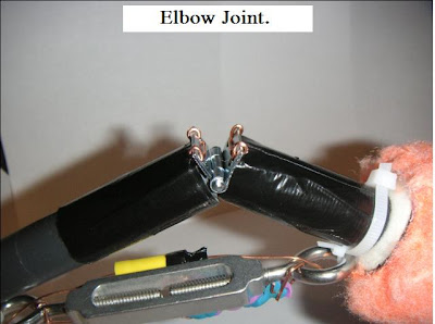
This photo shows the details of the elbow joint and how it allows for movement up and down but very little flexibility from side to side (that is by a rotating joint in the upper arm) This joint also shows there is a limit to the flexibility in the downward vertical plain, most peoples arm will not extend much further past straight and level.

In this photo you can see where my model first gets the sensory signal, this could be from touch of the finger sending a nerve impulse to the sensory neuron or something brushing up against the skin where a nerve signal is sent to the dendrites and then transmitted through the axon.

Here is the final picture of my movable limb. In this one the limb is rotated on its side to get a detailed shot as if you were looking at a top view from above the arm. The axon extends the full length of the arm and is the connecting point which allows the transmission of all signals to and from every part of the body to the spinal cord first.
Conclusion:
I thought this lab was challenging and very helpful in developing a better understanding of the nervous system. To take the concepts in the book and try to combine them in a visual model is very tricky, but does help to bring a deeper understanding of complexity in your arm. I know there are parts of my model which do not show some of the process which occur in a human arm, but my goal was to capture the major functions and I think I did that. One of the most important functions I wanted to capture was the actin-myosin unit and I think I designed an arm which shows how this process works. Overall someone who did not have an strong background in human biology could view my model and the lab write up and begin to grasp the basic mechanics of what makes an arm move on the microscopic level.
No comments:
Post a Comment